
Medical Imaging Technology
When it comes to diagnosing and treating spine related issues, being able to see beyond the scope of the naked eye is absolutely imperative. Luckily, as medical technology have advanced, doctors have several options available to help see what’s going on inside your body. Between X-rays, CT scans and MRIs, doctors and surgeons can create detailed images of the inner workings of your body that can be used to diagnose illnesses and abnormalities, recommend treatments, and even plan complex surgical procedures.
X-Rays
First discovered in 1895, the utilization of x-ray technology has certainly evolved over time. X-rays work by allowing low levels of electromagnetic radiation to pass through the body to create a 2D image of denser structures in the body. Different parts of the body absorb the energy from these rays differently, so while these rays may pass through soft tissue with ease they have a much harder time passing through bone. As a result, bones appear bright white on the image making x-rays ideal for diagnosing bone, teeth and joint issues within the body. X-rays can also help detect and diagnose abnormalities in the body such as tumors, pneumonia and even certain forms of cancer.
CT Scans
Computerized Axial Tomography, often referred to as a CT or CAT scan, uses specialized x-ray technology to create images of the body. CT scans produce 2-dimensional images of a “slice” or section of the body. These slices can be compiled and used to construct full 3D images. The CT scan can see hundreds of different levels of density as well as see tissues within a solid organ. A CT scan, in a sense, is like looking at one slice of bread at a time within a whole loaf. In order to get a more detailed image it may be necessary to use contrast dyes or ionizing radiation before the procedure. A CT scan can help provide doctors and surgeons with a detailed picture of a person’s tissues, organs, and skeletal structure.
MRIs
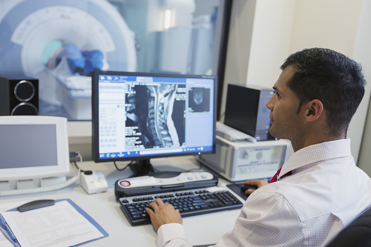
Magnetic Resonance Imaging, or MRI, is a test that utilizes powerful magnets, radio waves and sophisticated software to create a detailed, cross-sectional image of the inner workings of your body. The MRI machine creates a magnetic field inside of your body as radio waves are sent to the targeted area. This process causes the fat and water molecule protons in your bone and tissue to resonate, creating a computer-generated image. With an MRI, doctors can clearly view bones, joints, ligaments, blood vessels, cartilage and other soft tissue structures within the body. MRI’s are generally considered to be the most detailed imaging technology, able to help diagnose and treat soft tissue damage more accurately.
William Capicotto MD, PC
While there is no one-size-fits-all to diagnosing and treating back pain, improvements in technology continue to help give spine surgeons the resources and knowledge needed to treat a variety of conditions. As imaging technology continues to advance, it can continue to help make diagnosing and treating intricate structures of the body such as the spinal column more accurate and efficient, leading to shorter surgery times and quicker recoveries.
If you’re currently experiencing chronic, worsening, or debilitating back pain, don’t hesitate to contact our office to schedule your next visit. Simply call 716) 881-0382 today.