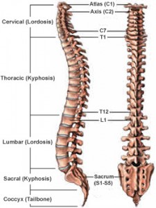
 Your spine is made of 33 bones stacked on top of each other. The spinal column provides the main support for your
body. The spinal column also protects the spinal cord from injury. An adult spine has a natural S-shaped curve when viewed from the side. These curves work to absorb shock, maintain balance and allow for a full range of motion throughout the spinal column. Muscles, ligaments, tendons, and nerves, along with the bones of your spinal column allow for you to stand upright, bend, and twist. Discover more about your spine by learning some basic spinal anatomy.
Muscles
Two major muscle groups affect your spine: the extensors and the flexors. The extensor muscles are attached to the back of the spine and allow for us to stand and lift objects. The flexor muscles are in front of the spine, including the abdominal muscles. They enable us to flex and bend forward.
Vertebrae
The 33 bones that interlock to form the spinal column are called vertebrae. The vertebrae are numbered and divided into 5 regions: cervical, thoracic, lumbar, sacrum, and coccyx. The vertebrae of the coccyx and sacrum are fused. As a result, only the top 24 bones are moveable.
Cervical
The cervical vertebrae are the vertebrae of your neck. These 7 bones (C1 to C7) work to support the weight of the head. C1 connects directly to the skull, allowing the head to nod. C2 has a projection called the odontoid, which C1 pivots around, allowing for side to side motion.
Thoracic
Twelve vertebrae (T1 to T12) make up the Thoracic spine. This section is in the mid back. It holds the rib cage, thus protecting the heart and lungs. There is a limited range of motion in the thoracic spine.
Lumbar (low back)
The lumbar spine is made up of 5 vertebrae (L1 to L5). These vertebrae are much larger in size. Their size allows them to absorb the stress of lifting and carrying heavy objects. It also helps to bear the weight of the body.
Sacrum
The sacrum is the next 5 vertebrae, which are fused together. They connect the spine to the hip bones. Along with the hip bones, they form a ring called the pelvic girdle.
Coccyx region
The last four vertebrae make up the coccyx region. Like the sacrum, they are also fused together. Also called the tailbone, the coccyx provides attachment for ligaments and muscles to the pelvic floor.
Intervertebral discs
An intervertebral disc separates each vertebra. The function of the discs is to cushion the bones, thus keeping them from rubbing together. The outer ring of the disc, the annulus, has bands that attach between the bodies of the vertebrae. Inside the disc is the nucleus, which is full of gel. The gel-filled nucleus is made of fluid which is absorbed at night when you are lying down and pushed out during the day as you move. The fibers of the annulus pull the vertebrae together against the nucleus. The vertebrae to roll over the gel of the nucleus, cushioning motion.
Facet Joints
Each vertebra has four facet joints: Two that connect to the vertebrae above and two the connect to the vertebrae below. The facet joints allow for back motion.
Ligaments
The ligaments are the strong bands that hold vertebrae together. They stabilize the spine, thus protecting the discs. There are three major ligaments of the spine. The anterior longitudinal ligament and posterior longitudinal ligament run from top to bottom and prevent excessive movement of the vertebral bones.The ligamentum flavum attaches between posterior arch of the vertebral bone.
Spinal cord
The spinal cord is about 18 inches long and the thickness of your thumb. It runs within the spinal canal from the brainstem to the 1st lumbar vertebrae. At the end of the spinal cord, the cord fibers separate and continue down the spinal canal to your tailbone, branching off to your legs and feet. In general, the main function of the spinal cord is to relay messages between the brain and the body. Messages for movement and sensation are sent from the brain through the cord. Reflexes are when the spine reacts without sending information to the brain in order to protect the body from harm. Damage to the spinal cord can result in a loss of sensation or motor function below the level of the injury. For example, you had an injury to your cervical area, sensory and motor loss can occur in your arms and legs (Tetraplegia).
Spinal nerves
Thirty-one pairs of spinal nerves branch off the spinal cord. These nerves assist in the relay of information between the spinal cord and the brain. Each spinal nerve has two roots. The ventral (front) root carries information from the brain. Its opposite, the dorsal (back) root, carries information to the brain. The nerve travels alongside the chord until it reaches its exit point. After it exits the vertebrae, the nerve branches off to the skin and muscles in the front and back of the body.


